Planktic foraminifera culturing on Green Island, Taiwan: Foram proxy development and genetics
|
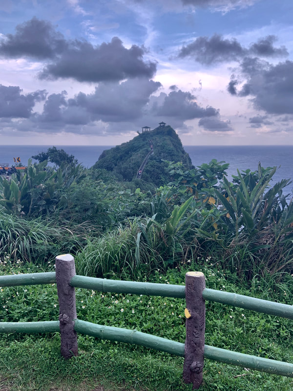
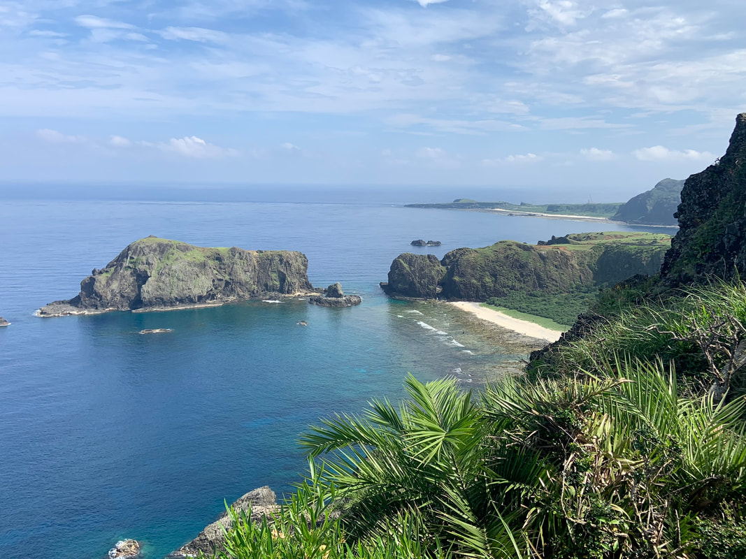
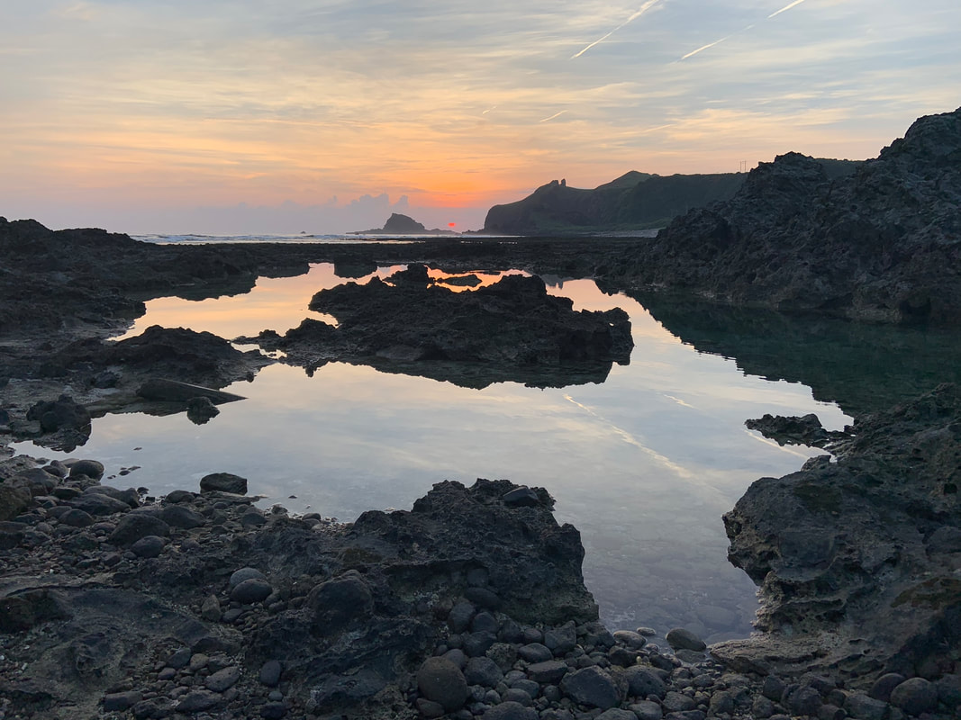
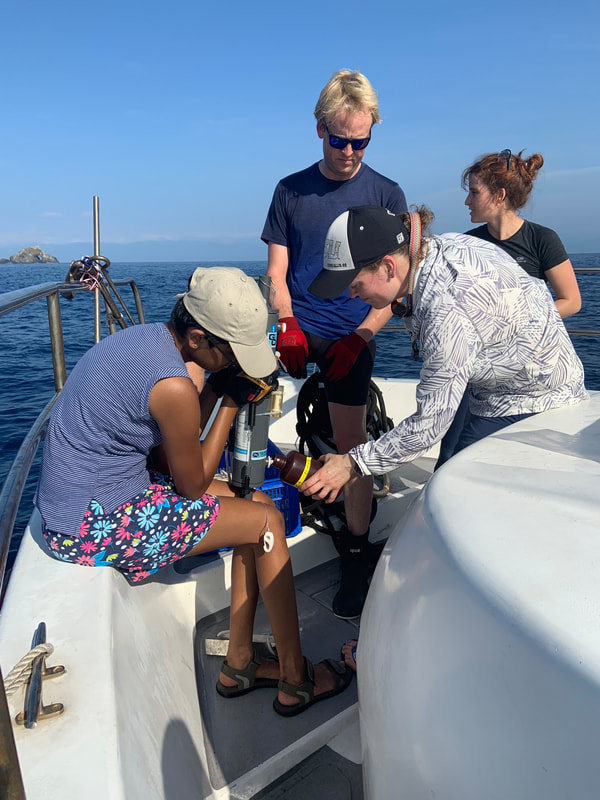
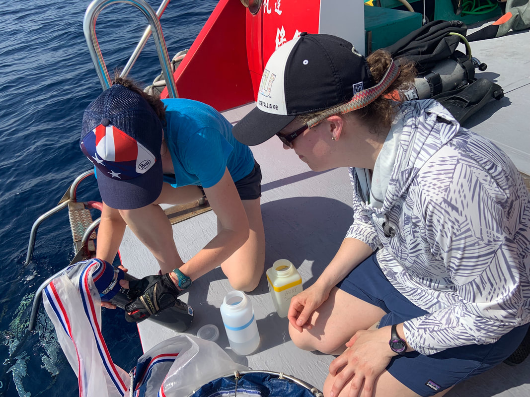
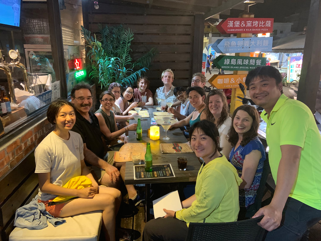
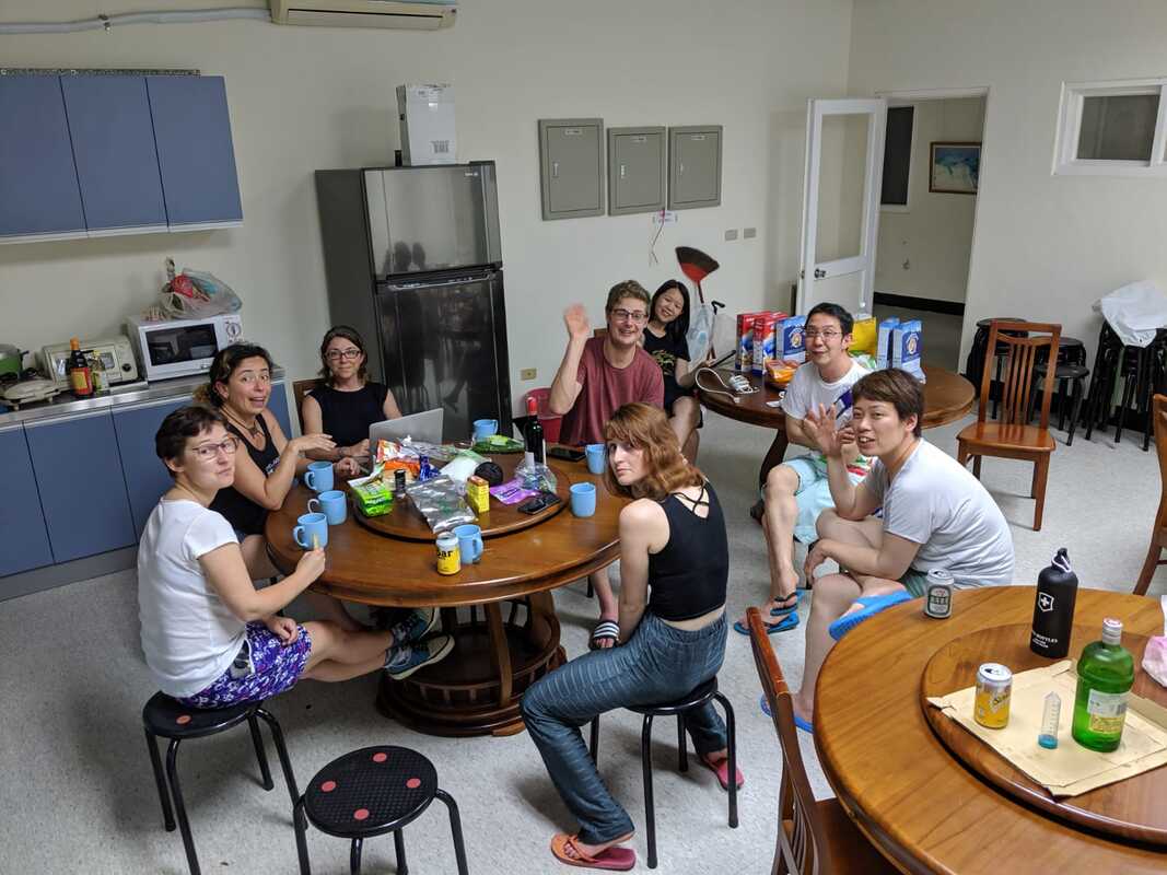
In May, 2019 we traveled to Green Island, Taiwan to culture planktic foraminifera at the Green Island Marine Research Station. The culture season was organized by Dr. Abby (Haojia) Ren, who is a professor at the National Taiwan University. The field season included researchers from NTU, Cambridge, UC Davis, Oregon State University, University of Angers, JAMSTEC, U of Arizona, and more. It was an exciting field season because it was the first time any of us had attempted a field season at this location and we didn't know what to expect. It was also the most difficult, in terms of logistics (getting there required planes, trains, taxis, shuttles and a ferry).
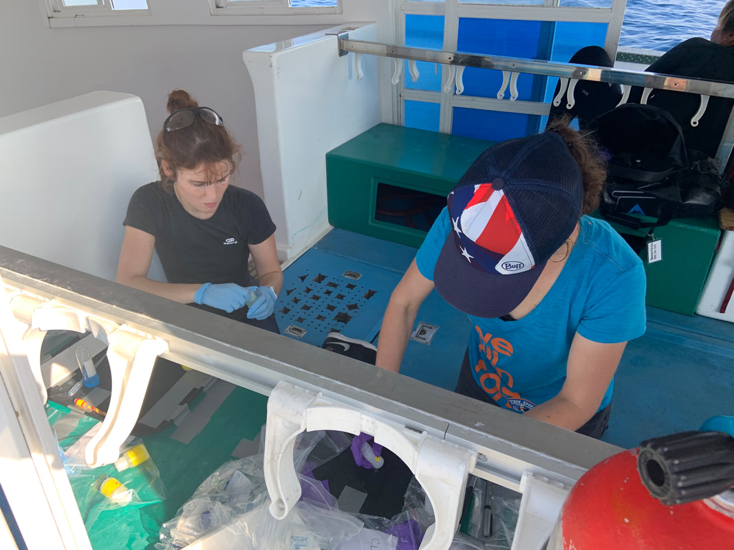
The research goals for my group included collecting non-spinose foraminifera to expand our research beyond the neogloboquadrinid species and collect specimens for genotyping and metabarcoding (using 16S and 18S metabarcoding we plan to assess symbiotic and microbial associations). We also conducted a few side experiments to track the uptake of S, C, and N. And light manipulation experiments with spinose forams. We had a second field season planned for 2020, but due to the pandemic, the field season was cancelled. I hope to head back to Green Island in 2024. Some photographs from our field season are below. Learn about our Catalina field projects HERE. Learn about our Coastal Oregon projects HERE. |
Select projects:
|
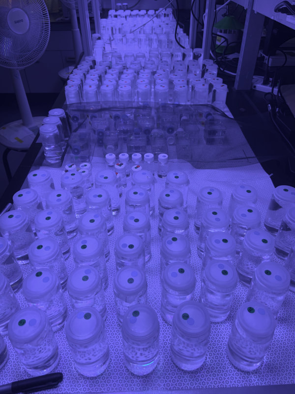
So. Many. Forams. Many of the experiments being conducted didn't require temperature control so the culture jars were kept on a table in the lab. There were well over a hundred specimens in culture at any given time. The blue light 'hue' is from the reef lights we grow the forams under, which doesn't photograph well. True color is not quite as blue!
|
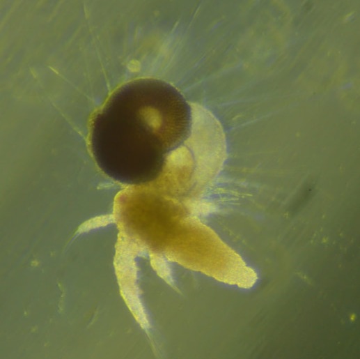
This P. obliquiloculata was fed a single frozen (thawed) 1-day old artemia brine shrimp. Click on the image for a larger version where you can see the new chamber (no cytoplasm in it yet) and the rhizopodia.
|
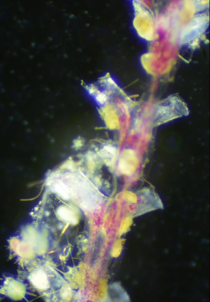
The brown specs in this photograph are tiny (100 micron) juvenile G. menardii that are attached to what we thing might be part of a branched bryozoan. The forams were literally stuck like glue and we couldn't get them dislodged. We think non-spinose species try to find substrate to adhere to in the water column.
|