Electron microprobe images of planktonic foraminifera Mg/Ca variability
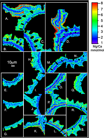
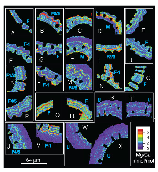
As part of my PhD research, I used electron microprobe imaging to map the variability of Mg in the shells of several species of planktic foraminifera. In order to generate data using this method, the tiny (a few hundred microns across) foram shells are embedded in epoxy and sanded to reveal the cross-section of the shell. The surfaces are then polished using diamond paste or diamond lapping sheets, and finally carbon coated. The electron beam can be rastered across the thin section and once the data is processed you are left with a beautiful image of the Mg/Ca variability within the shell. I generated image maps of fossil specimens of G. ruber, G. sacculifer, and N. dutertrei to explore Mg-variability in well preserved (shallow core) and poorly preserved (deep core) samples. Mg/Ca variability was also compared in modern and LGM specimens to compare Mg/Ca ratios in specimens that formed in warmer (modern) vs. cooler (LGM) conditions. Preliminary results were published in 2010. We used this technique to assess changes in shell chemistry that occur when the shells dissolve on the seafloor (results published in Paleoceanography). Advancements in microprobe imaging techniques (and instruments) yield much higher resolution images. (Figure from Fehrenbacher and Martin, 2014)
Relevant Publications: Fehrenbacher, J.S., P.A. Martin, (2014) Exploring the dissolution effect on the intratest Mg/Ca variability in the planktonic foraminifera Globigerinoides ruber, Paleoceanography, doi/10.1002/2013PA002571 Fehrenbacher, J.S., P.A. Martin (2010), A comparison of intrashell Mg/Ca variability of the planktonic foraminifera G. ruber, G. sacculifer, and N. dutertrei determined by electron microprobe image mapping, 2010 IOP Conf. Ser.: Earth Environ. Sci. doi:10.1088/1755-1315/9/1/012018 |
Select projects
|
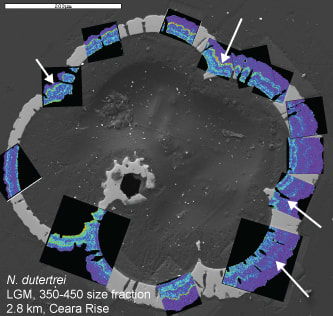
Data was also generated for the species N. dutertrei: (unpublished)