|
And I'm not kidding either... Case in point: This is a picture of an O. universa. We caught this lovely specimen this past summer on Catalina Island. You can clearly see its thin calcite spines, symbionts around the sphere, and juvenile inside the recently formed adult sphere. This little critter is less than a millimeter wide... with spines perhaps 4 mm. This species is quite photogenic. Really. It has even adorned the cover of Science. 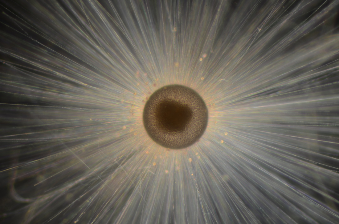 Here is another beauty: this one was caught in a plankton net. It has broken spines (which later re-grew) because the plankton net often beats the forams up a bit. Even those with no spines can recover. This one had a very new final chamber and you can clearly see the juvenile trochospiral shell on the inside and the cytoplasm that is starting to infill the final sphere. 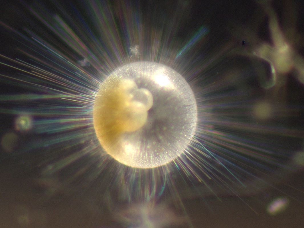 Other foraminifer species are equally beautiful. Especially those deep dwellers... I admit, I'm biased because I've spent a LONG time studying them. Mostly, I have used their geochemistry to infer changes in the earth's climate. More recently, however, I've delved into understanding how they tick... especially these lesser known deep dwelling species that are more difficult to study in laboratory conditions. I guess I like a challenge. The deep dwellers are really quite beautiful too. Take a look below... Really. Go. Look. You won't be disappointed. I'll upload more photos soon. 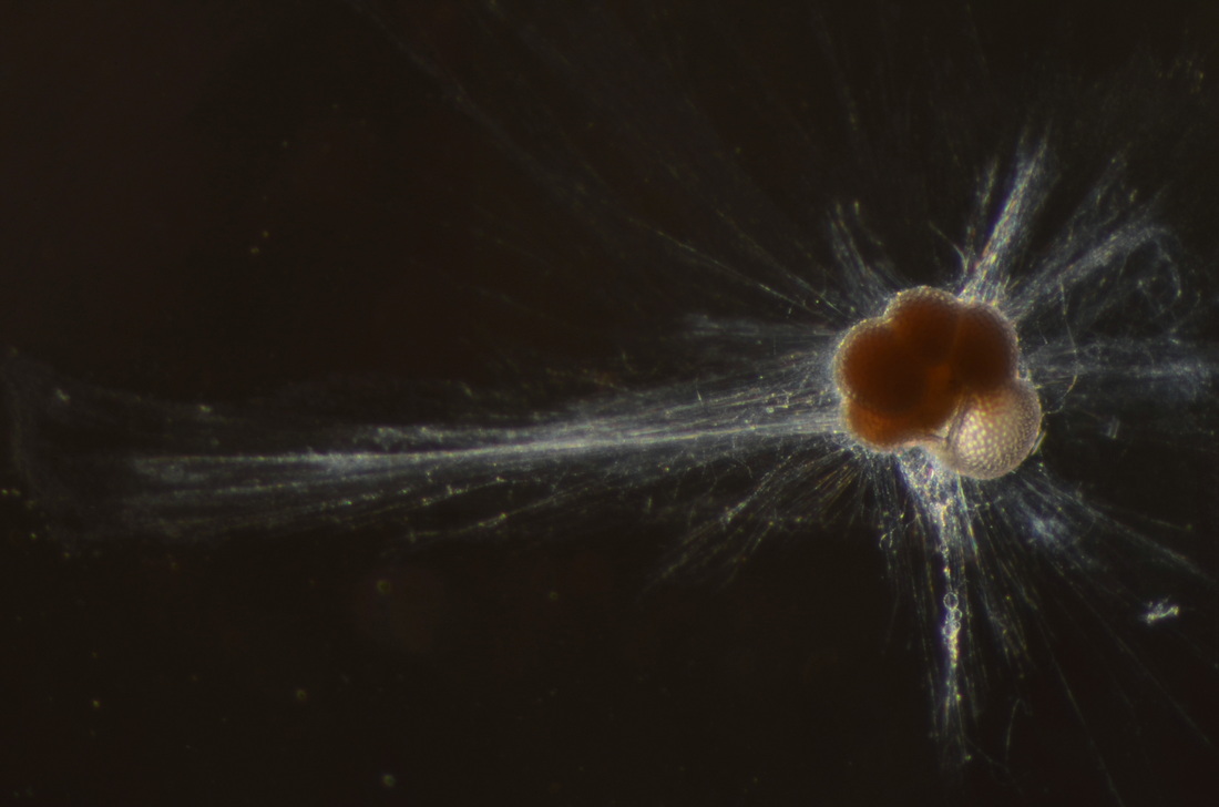 This is an N. dutertrei with a new chamber that is infilling with cytoplasm. This specimen formed this chamber in the ocean, then finished calcifying while in culture. The 'web' around he foram it the rhizopodial/pseudopodial network. 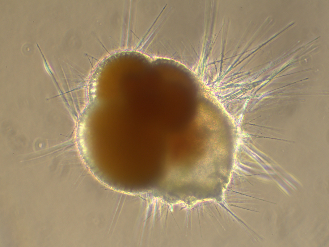 This foram was captured with a brand new chamber. The chamber wall was so thin that when the foram 'went benthic' within the culture jar, the wall of the new chamber 'stuck' to the jar and was pulled into a deformed shape when the foram tried to move. This is exactly how the final chamber formed, only it added about 20-25 microns of calcite in culture. It has a very thick crust and the final chamber was shaped exactly like you see here... 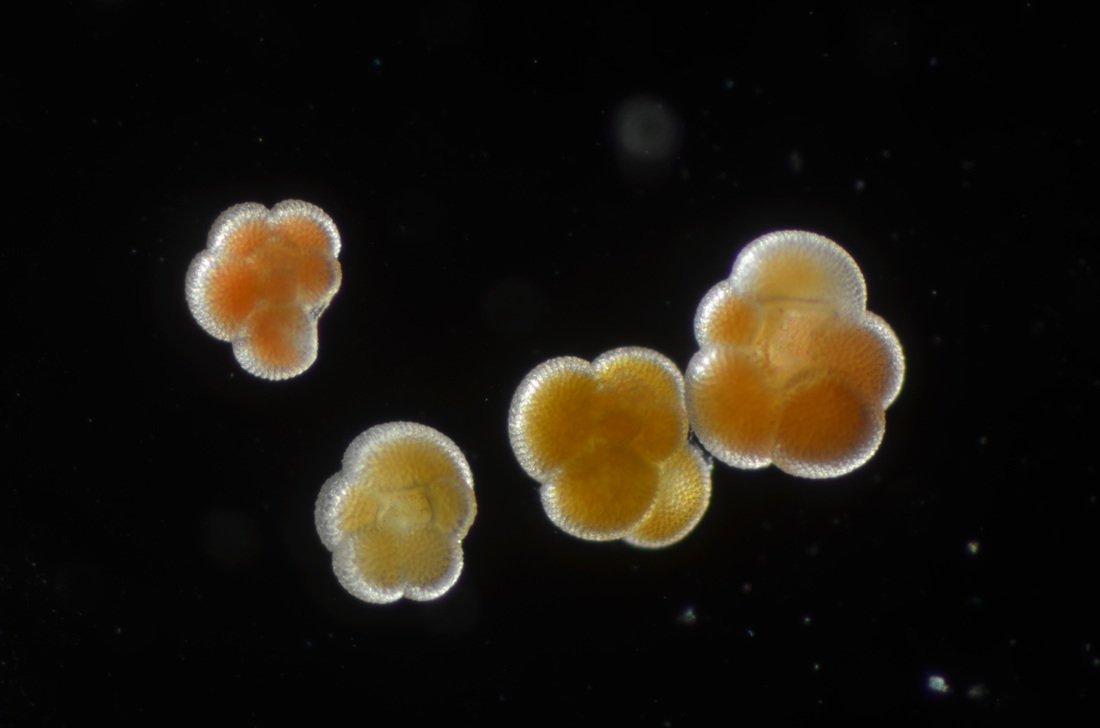 These are four N. dutertrei in different stages of ontogeny. The upper left specimen would have likely added another chamber. The second from the left would have been a very large specimen had it been given a chance to add more chambers. All four specimens were photographed, transferred into small vials, and then fixed in glutaraldehyde/paraformaldehyde for TEM analysis. 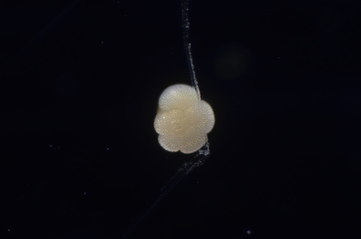 A gametogenic N. dutertrei. These species 'bleach' before they release gametes. A day or two after this happens, gametes are released and the shell is left empty.
57 Comments
|
 RSS Feed